
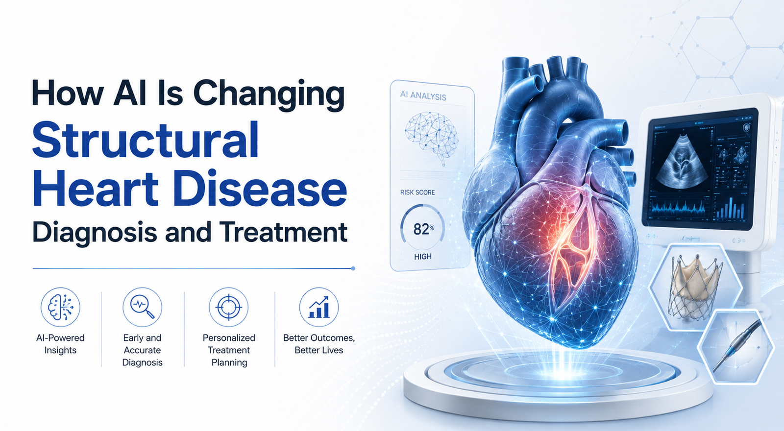
Structural heart disease (SHD) — conditions affecting the heart’s valves, chambers, walls, and muscle — is one of the most underdiagnosed categories in cardiovascular medicine. Millions of people live with undetected valve disease, cardiomyopathy, or reduced heart function simply because the tools needed to catch it, like echocardiography, require specialized equipment and trained interpreters that aren’t available everywhere. Artificial intelligence is closing that gap, and 2026 has become something of an inflection point: AI tools are moving out of research papers and into emergency departments, cath labs, and community clinics.
The Diagnostic Bottleneck AI Is Solving
Echocardiograms remain the gold standard for confirming most forms of structural heart disease, but they’re expensive, resource-intensive, and dependent on specialist availability. Electrocardiograms (ECGs), by contrast, are cheap, fast, and performed hundreds of millions of times a year worldwide. The core insight driving much of today’s AI innovation in structural heart disease is that a standard 12-lead ECG contains far more diagnostic information than the human eye can extract — and deep learning models can read those hidden signals.
The best-known example is EchoNext, a deep learning model developed by researchers at Columbia University Irving Medical Center and trained on more than a million paired ECG and echocardiogram records. In head-to-head testing against thirteen cardiologists reading the same ECGs, <cite index=”16-1″>EchoNext correctly identified structural heart problems 77% of the time, compared with 64% accuracy for the cardiologists</cite>. The tool has since received FDA clearance and can flag <cite index=”17-1″>six categories of structural heart disease, including valve problems and impaired pumping function, directly from a routine EKG</cite>.
This matters because early detection changes outcomes. Research cited in the EchoNext validation studies found that in a survey of adults over 65, screening turned up nearly as many new cases of valvular heart disease as had previously been diagnosed, <cite index=”14-1″>more than doubling the known prevalence of valvular disease in that population once undiagnosed cases were counted</cite>.
From Screening to Real-World Clinical Impact
What sets this current wave of AI apart from earlier hype cycles is documented downstream impact — cases where an algorithm’s flag led directly to a life-changing intervention. Columbia researchers have published what they describe as the first documented case of an AI-ECG algorithm detecting undiagnosed severe aortic stenosis that led straight to a transcatheter aortic valve replacement (TAVR). Similar case reports describe AI flagging severe mitral regurgitation that led to valve repair, and one instance where AI-assisted diagnosis contributed to a patient receiving a heart transplant.
Hospitals are also testing “silent deployment” of these models — running them quietly in the background without altering clinical workflow, just to measure impact. In one such study presented at THT 2026, researchers found that <cite index=”3-1″>nearly half of patients flagged as high risk for structural heart disease would not have been referred for an echocardiogram under standard care alone</cite>. That’s a striking number: it suggests AI isn’t just replicating what clinicians already catch — it’s surfacing an entirely separate population of at-risk patients.
AI’s Growing Role in Imaging and Procedural Planning
Diagnosis is only half the story. Once structural heart disease is identified, clinicians increasingly rely on AI to plan and support treatment, particularly for minimally invasive transcatheter procedures like TAVR and transcatheter mitral or tricuspid repair. A recent review in the Journal of the Society for Cardiovascular Angiography & Interventions found that AI tools can now automate the quantification of cardiac anatomy and hemodynamics with a level of consistency that outperforms traditional manual measurement, supporting more precise device sizing and procedural planning for complex structural interventions.
Cardiac CT is another area where AI has become deeply embedded. According to cardiac imaging specialists speaking ahead of SCCT’s 2026 annual meeting, AI now touches nearly every stage of cardiac CT, <cite index=”2-1″>from image acquisition and reconstruction to automated plaque quantification, workflow optimization, and advanced physiologic assessment</cite>. That expanding footprint is helping cardiologists move from reactive treatment toward earlier, biology-driven prevention strategies.
Why This Moment Feels Different
Cardiology has seen waves of AI enthusiasm before, but clinicians now point to a genuine shift from research promise to clinical practice. As one cardiology AI researcher put it in a recent American College of Cardiology feature, the field has moved from speculative pilot studies to tools embedded in real clinical workflows, complete with FDA clearances and deployment across hospital systems. Randomized controlled trials — including Columbia’s SAGE trial and Montreal Heart Institute’s HEART-AI study — are now underway to formally measure whether AI-guided screening changes management and outcomes at scale, not just in retrospective analysis.
Considerations for Clinical Adoption
None of this eliminates the clinician from the equation — if anything, current evidence suggests AI performs best as a triage and decision-support layer rather than a stand-alone diagnostician. Reviewers have flagged real challenges ahead: ensuring model accuracy holds up across diverse patient populations, addressing clinician trust and adoption, managing the cost and accessibility of follow-up imaging once a patient screens positive, and building explainability into models so physicians understand why a given prediction was made.
For device and therapy manufacturers in the structural heart space, this shift has practical implications too — AI-assisted anatomical assessment and procedural planning tools are increasingly factoring into how care teams select and size interventional devices, which makes accurate, well-validated imaging data more important than ever.
The Bottom Line
AI isn’t replacing cardiologists, but it is fundamentally expanding who gets screened, how early disease is caught, and how precisely procedures are planned. With FDA-cleared tools already reaching hundreds of thousands of clinicians and multiple randomized trials underway, structural heart disease is emerging as one of the clearest proof points for AI’s value in modern cardiovascular care.
Frequently Asked Questions
Can AI diagnose structural heart disease from a standard ECG? Yes. AI models such as EchoNext are trained to detect signals in a routine 12-lead ECG that indicate a likelihood of structural heart disease, including valve disease and reduced heart function, and then recommend follow-up echocardiography for confirmation.
Is AI used to guide structural heart procedures like TAVR? Increasingly, yes. AI-assisted imaging analysis helps clinicians quantify cardiac anatomy and hemodynamics more precisely, supporting device selection and procedural planning for transcatheter valve interventions.
Does AI replace the need for a cardiologist? No. Current tools function as screening and decision-support aids. Diagnosis confirmation and treatment decisions still involve cardiologist review and standard imaging like echocardiography.